Why Ultrasound Matters in Modern Pet Care
Pets can’t tell us when something feels wrong, so subtle changes in behavior or appetite often leave families guessing. Maybe your dog suddenly stops eating, or your cat begins hiding and losing weight without explanation. Instead of waiting and worrying, ultrasound gives veterinarians a safe, real-time way to look inside the body without surgery. This simple, noninvasive test helps detect illness earlier, rule out hidden problems, and guide the right treatment before conditions become more serious.
At Santa Monica Veterinary Group, ultrasound isn’t just a diagnostic tool. It’s part of a bigger commitment to providing thorough, compassionate care. By combining imaging with preventive services, urgent care, and advanced treatments, we ensure that pets get more than a diagnosis. They get a clear plan, tailored to their needs, delivered with comfort and precision.
Understanding Ultrasound in Veterinary Medicine
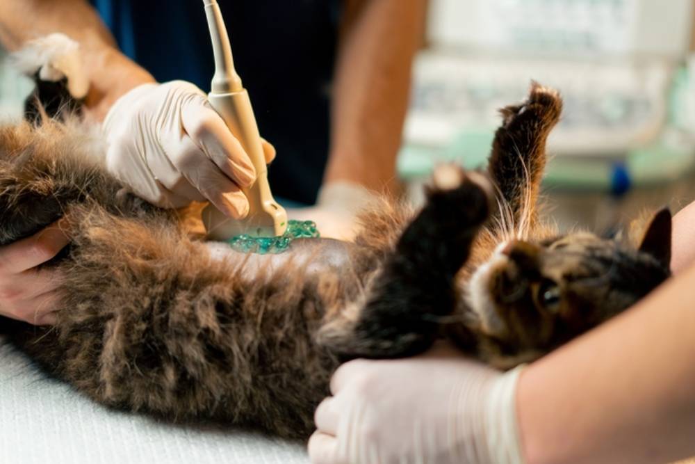
Ultrasound imaging uses sound waves to create live pictures of internal organs. Unlike X-rays, which are best for bones, ultrasound excels at soft tissue structures such as the liver, spleen, heart, kidneys, and intestines.
Most dogs and cats don’t require sedation for this exam, though mild sedation may be recommended if a pet is very anxious or painful. Because it is painless, versatile, and safe, ultrasound has become one of the most trusted diagnostic tools in veterinary medicine.
When Vets Turn to Ultrasound for Answers
Ultrasound is often recommended when a pet shows persistent or unexplained symptoms:
- Cats with long-term vomiting or diarrhea
- Dogs with ongoing weight loss or chronic diarrhea
- Pets with new heart murmurs or coughing at rest
- Abdominal swelling, weakness, or sudden loss of energy
Because many of these conditions share overlapping signs, ultrasound helps narrow the cause quickly. For urgent concerns, our hospital also offers same-day urgent care so pets can be evaluated without delay.
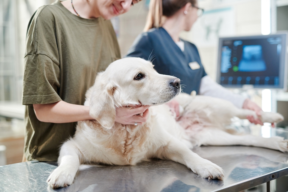
Abdominal Ultrasound: Digestion, Kidneys, and More
Abdominal scans are among the most common types of ultrasound we perform. They can reveal:
- Digestive tract problems: Inflammatory changes from IBD in cats, thickened intestines caused by lymphoma, or blockages from foreign objects.
- Liver and spleen issues: Chronic disease, cirrhosis, or tumors such as hemangiosarcoma in dogs. Ultrasound can also confirm hemoabdomen, a life-threatening bleed into the abdomen.
- Kidneys and urinary tract: Detecting stones, obstructions, or inherited conditions like polycystic kidney disease in cats. Guided cystocentesis may be used for sterile urine collection.
- Gallbladder and bile ducts: Ultrasound can spot gallstones, sludge, inflammation, or bile duct obstruction, which may explain vomiting, jaundice, or abdominal pain.
- Stomach and pancreas: Changes in stomach wall thickness or pancreatic inflammation can help diagnose gastritis or pancreatitis.
- Lymph nodes: Enlarged abdominal lymph nodes can indicate infection, inflammation, or cancer. Measuring and monitoring them over time helps track disease progression or response to treatment.
Ultrasound is also a standard tool in cancer screening, where it helps identify metastasis or subtle organ changes long before external symptoms appear.
Heart Health: The Role of Echocardiograms
When a pet develops a new murmur or irregular heartbeat, an echocardiogram (cardiac ultrasound) is often the next step. An echocardiogram shows the heart’s chambers, valves, and blood flow in real time. It can identify congenital defects, cardiomyopathy, pericardial effusion, and even heartworm complications.
Early detection through echocardiography helps veterinarians design treatment plans that slow disease progression and improve quality of life.
Reproductive and Uterine Imaging
Ultrasound is invaluable in evaluating reproductive health. For female pets, scans can confirm pregnancy, assess fetal viability, and estimate litter size. Just as importantly, ultrasound can detect uterine infections like pyometra, which is a life-threatening emergency requiring surgery. Identifying these conditions early ensures faster, safer intervention.
Lung Ultrasound: Beyond X-Rays
While chest X-rays are commonly used for respiratory issues, ultrasound has unique advantages. It can help identify fluid buildup in or around the lungs, guide thoracocentesis (removal of fluid), and highlight lesions or tumors near the chest wall. For pets in respiratory distress, lung ultrasound is often combined with other imaging to reach a clear diagnosis quickly.
Ocular Ultrasound: Seeing the Eyes Clearly
In cases where the eye is too opaque to examine with standard tools- such as severe cataracts, bleeding, or trauma- ocular ultrasound becomes essential. It allows veterinarians to evaluate the retina, lens, and other internal structures, helping diagnose retinal detachment, tumors, or lens luxation. This technology is especially important for guiding treatment when vision-threatening conditions are suspected.
Ultrasound-Guided Biopsies and Sampling
Sometimes, pictures alone are not enough to reach a diagnosis. Ultrasound can be used to guide fine-needle aspirates or biopsies, which involve collecting a small sample of cells or tissue from an abnormal area seen on the scan.
- A fine-needle aspirate uses a thin needle to withdraw just a few cells. It is quick, often requires little to no sedation, and can provide information about whether a mass is likely inflammatory, infectious, or cancerous.
- A biopsy collects a slightly larger piece of tissue, giving pathologists a more detailed look at the structure of the cells. This helps confirm the exact type of disease and how aggressive it may be.
The samples are usually sent to a laboratory, where they are examined under a microscope. Results can reveal whether an organ change is due to cancer, infection, or another condition, allowing veterinarians to create a more precise treatment plan.
This minimally invasive approach reduces the need for exploratory surgery, speeds up answers, and helps families make informed decisions about next steps.
FAST Scans: Ultrasound in Emergencies
In critical situations, ultrasound can save lives. FAST (Focused Assessment with Sonography for Trauma) scans are rapid exams that detect free fluid in the chest or abdomen, ruptured organs, or internal bleeding. If a pet collapses, is struck by a car, or has severe breathing trouble, a FAST scan often provides the first crucial answers.
For emergencies during business hours, our team is equipped to handle urgent cases immediately. Visit our urgent care page for guidance if your pet experiences sudden distress.
From Imaging to Action: The Next Steps
Ultrasound rarely stands alone. Results are often combined with bloodwork, X-rays, CT, MRI, or endoscopy to build a complete picture of your pet’s health. Some conditions can be managed with medication and diet, while others may require surgical solutions.
Our team walks families through every result, outlining clear treatment paths that fit each pet’s needs and lifestyle.
Comprehensive Veterinary Ultrasound in Santa Monica
From subtle digestive problems to life-threatening emergencies, ultrasound remains one of the most versatile tools in modern veterinary medicine. It offers clarity without invasive surgery, helps guide treatment, and supports long-term monitoring of chronic conditions.
At Santa Monica Veterinary Group, we integrate advanced ultrasound services into a full spectrum of wellness, urgent care, and specialty treatments. If your pet is showing concerning symptoms, contact us today or request an appointment online. With thorough diagnostics, compassionate care, and a focus on your pet’s comfort, we deliver more than answers- we deliver a better way to care.


Leave A Comment