
Fine Needle Aspirate vs. Biopsy: How Your Vet Decides Which Test Your Pet Needs
You were petting your dog and your hand stopped on something that was not there last week. Or you noticed a small bump on your cat while she was sitting on your lap. The first thought is almost always the same: is this cancer? The honest answer is that no one can tell from the outside, no matter how experienced. Some malignant tumors feel soft and squishy. Some entirely benign masses feel firm and fixed. The only way to get a real answer is diagnostic testing, and the two main tools we use are fine needle aspirate (FNA, where a thin needle collects cells from the mass for microscopic evaluation) and biopsy (where a tissue sample is sent to a pathologist for a more detailed analysis).
The encouraging news: most lumps are not emergencies, and many turn out to be benign. The catch is that you cannot know which is which without testing. At Santa Monica Veterinary Group, we evaluate lumps and masses every day and tailor the diagnostic approach to each pet’s situation. Our in-house diagnostic capabilities and surgical options, including CO2 laser surgery for precise mass removal, allow us to move quickly from discovery to diagnosis to treatment when needed. If you have found a new lump on your pet or noticed a change in an existing one, call us or request an appointment so we can take a closer look.
Why You Cannot Diagnose a Lump by Looking at It
Signs of cancer in pets do not correspond reliably to a mass’s appearance. Malignant tumors can feel soft and moveable, just like a fatty lump. Benign masses can feel firm and fixed, just like something more concerning. Surface texture, growth rate, color, and location all give us clinical clues, but none of them give a diagnosis.
Cancer in pets affects roughly one in four dogs over their lifetime, and rates climb significantly in senior pets. That is common enough that testing any new lump is routine practice rather than overreaction. The earlier we get an answer, the more treatment options are on the table, and the better the outcomes tend to be.
Fine Needle Aspiration: The First Diagnostic Step
How Fine Needle Aspiration Works
A fine needle aspiration inserts a thin needle into the mass to collect cells or fluid, which are spread on a slide and examined under a microscope. The procedure takes a few minutes. Most pets tolerate it about the same as they do a vaccination, no anesthesia or sedation needed. We can sample multiple areas of a mass or several different masses in a single visit, and results from our reference laboratory typically come back within a few business days.
What FNA Can and Cannot Diagnose
Cytology examines individual cells rather than the surrounding tissue structure, and that distinction matters for what FNA can definitively answer. Skin cytology is especially informative for superficial lesions and skin surface changes.
Conditions that FNA reliably diagnoses include lipomas, mast cell tumors, cysts, reactive vs. malignant lymph nodes, and several round cell tumors like histiocytomas and plasmacytomas. For these, the cells under the microscope are distinctive enough that the diagnosis is clear without further testing.
Where FNA falls short is with tumors that release cells poorly into a needle. Fibrosarcomas, poorly exfoliating mammary tumors, and certain other sarcomas may produce non-diagnostic samples even when the mass is malignant. A non-diagnostic FNA does not rule out cancer; it just means the test could not get enough information to commit to an answer. For a growing or suspicious mass with a non-diagnostic FNA, biopsy is the appropriate next step rather than continued monitoring.
Skin Masses That Look Similar but Behave Very Differently
One of the main reasons cytology matters so much is that several common skin masses can look nearly identical on the outside but have completely different prognoses. Telling them apart by appearance alone is not reliable, even for experienced veterinarians.
Commonly Confused Skin Masses in Dogs
Skin cancer in dogs comes in at least five distinct forms, and several of them can look similar to common benign masses. The most frequently confused include:
- Lipomas: benign fatty masses that are extremely common in middle-aged and older dogs. They typically feel soft, moveable, and sit just under the skin. The catch is that they can feel identical to certain malignant tumors on physical exam, which is why even a classic-feeling lipoma is worth aspirating at least once.
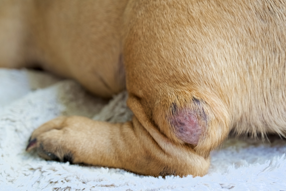
- Mast cell tumors: the most common skin cancer in dogs, and one of the best reasons never to skip testing a lump. They can look like just about anything, from a soft fatty-feeling bump to a red, angry-looking swelling, and they can change size from day to day. Cytology usually makes the diagnosis quickly, and grade and stage guide treatment from there.
- Histiocytomas: benign, fast-growing, button-like masses that typically appear in young dogs. Many resolve on their own, but they often look alarming enough that families assume the worst. Cytology confirms the diagnosis in minutes.
- Melanocytic tumors: range from entirely benign pigmented growths to aggressive malignant melanomas, depending on location and cell characteristics. Oral melanomas are particularly aggressive and need fast, thorough evaluation.
- Cysts: benign fluid-filled or keratin-filled sacs that can feel firm, soft, or in-between. Sebaceous and follicular cysts are common and usually harmless, but some “cysts” turn out to be something else once cytology is done.
Commonly Confused Skin Masses in Cats
Skin masses in cats deserve their own conversation because cats have a higher proportion of malignant masses than dogs. A lump that might be a routine fatty tumor in a Labrador needs more careful evaluation in a cat.
- Feline basal cell carcinoma: one of the most common skin tumors in cats, often appearing as a raised, firm, or ulcerated mass. Typically slow-growing and often curable with complete surgical removal, but needs diagnosis first to distinguish from more aggressive tumors.
- Squamous cell carcinoma: often starts as a small scab or crusty area that keeps coming back, progresses to ulceration, and can become locally invasive if not caught early. These are especially important to evaluate in white or light-colored cats. Any skin lesion that has not healed within a few weeks warrants evaluation, not a wait-and-see approach.
- Fibrosarcoma in cats: firm, locally aggressive tumors that most often develop in the skin and underlying tissue, sometimes at vaccine or injection sites. They tend to feel deep-seated and fixed rather than soft and moveable, and they are notorious for coming back if not removed with wide surgical margins. Biopsy is typically needed because these tumors often do not shed cells well on FNA.
- Feline mast cell tumors: behave differently in cats than in dogs. They often appear as raised, hairless, pink or reddish bumps on the head, neck, or trunk, and many of the cutaneous forms in cats are benign or behave less aggressively than the canine version. Cats also develop a visceral form involving the spleen or intestines, which is why any confirmed feline mast cell tumor warrants a broader workup.
Biopsy: When Tissue Architecture Is Needed
Types of Biopsy and How They Are Performed
A biopsy collects tissue rather than individual cells, which lets the pathologist evaluate cellular organization, invasion patterns, and structural relationships that simply do not show up on cytology slides.
The main biopsy approaches:
- Punch biopsy: a circular cutter removes a tissue cylinder; used for well-defined superficial masses
- Incisional biopsy: a wedge of tissue is removed without taking the whole mass; used when diagnosis is needed before planning definitive surgery
- Excisional biopsy: the entire mass is removed and submitted; combines treatment and diagnosis when the mass is small and surgically accessible
- Needle core biopsy: a larger-gauge needle collects a tissue core that preserves architecture without open surgery
All biopsy procedures require sedation or anesthesia for patient comfort and accurate sampling. Tumor diagnosis through histopathology is the diagnostic gold standard for cancer because it provides information that no other test can.
What Histopathology Reveals
Histopathology evaluates several things at once:
- Tumor type: the specific cancer confirmed at the tissue level
- Grade: how aggressive the cells look and how they are likely to behave
- Invasion: whether the tumor is infiltrating surrounding structures
- Margins: whether surgically removed tissue has clear edges or tumor cells right at the cut margin
Margin assessment after excision is one of the most clinically important pieces of information we can get. Without it, there is no reliable way to know whether the surgery was curative or whether more treatment is needed. Two tumors that look identical on FNA may behave completely differently and require different treatments, and histopathology resolves that ambiguity.
When Biopsy Is the Only Way to Get a Reliable Answer
Several clinical situations highlight why biopsy is sometimes the only path to a definitive diagnosis. Two come up often enough to be worth knowing about specifically.
Chronic GI Disease in Cats: IBD or Lymphoma?
Cats with chronic vomiting, diarrhea, and weight loss commonly have one of two conditions: inflammatory bowel disease in cats or feline lymphoma. These two conditions can produce nearly identical clinical signs, nearly identical bloodwork findings, and nearly identical ultrasound appearances of a thickened intestinal wall. They are also related, because untreated IBD can progress to lymphoma over time.
The treatments, however, are completely different. IBD is managed with diet changes, immunosuppressants, and sometimes antibiotics. Lymphoma requires chemotherapy. Guessing wrong means either a cat with cancer who is not receiving effective treatment, or a cat with IBD receiving chemotherapy they do not need.
Splenic Masses: Benign or Hemangiosarcoma?
When imaging finds a mass on the spleen, families often hear a frightening term: hemangiosarcoma. The concern is reasonable because hemangiosarcoma in dogs is aggressive and common in certain breeds, particularly Golden Retrievers and German Shepherds. But not every splenic mass is hemangiosarcoma. Roughly half of splenic masses turn out to be benign (such as a hematoma or nodular hyperplasia) and the other half malignant. The two types can look identical on ultrasound, and FNA of the spleen carries bleeding risk and often produces non-diagnostic results because these masses are full of blood.
Histopathology after splenectomy (surgical removal of the spleen) is typically the definitive answer. This is why the standard approach for most actively bleeding or large splenic masses is surgery first, with the full spleen sent to the pathologist. A benign result gives families relief and a cure. A malignant result guides decisions about further staging and treatment.
CO2 Laser Surgery for Mass Removal
Santa Monica Veterinary Group offers CO2 laser surgery for mass removals. Laser surgery provides precise tissue cutting that minimizes bleeding, reduces post-operative swelling, and causes less thermal damage to surrounding tissue than conventional electrocautery. For masses in sensitive locations, near critical structures, or where clean margins and minimal trauma matter most, CO2 laser surgery produces better outcomes than scalpel alone.
Choosing Between FNA and Biopsy
FNA is typically the first choice because it is fast, low-cost, and requires no anesthesia. Biopsy follows when:
- FNA returns non-diagnostic or inconclusive for a suspicious mass
- The tumor type requires grading for treatment planning
- Margin assessment after excision is clinically important
- Clinical suspicion for malignancy is high despite a benign FNA result
- The mass location makes FNA yield poor
The decision is rarely either-or. Many cases start with FNA and progress to biopsy based on what the cytology shows. The goal is always getting the right answer with the least invasive approach that will reliably provide it.
Monitoring Benign Masses Over Time
A benign result does not end the conversation. Masses can change over months to years, and a mass that aspirated as a clear lipoma this year can grow large enough to cause problems, develop a different character, or have new masses appear right next to it that look similar from the outside but are not.
A simple monitoring routine for any mass, even one with a benign diagnosis:
- Measure the mass. Use a ruler or calipers to note length, width, and approximate height. Photographs with a ruler next to the mass are also helpful for tracking.
- Write it down. A running list with the date, location, and size of each mass gives you and our team real data to compare over time.
- Check every month or two. A quick feel-over during a grooming session or belly rub is usually enough.
- Call us if anything changes. New color, new firmness, rapid growth, sudden size increase, ulceration, bleeding, or pain all warrant a recheck.
- Consider periodic re-aspiration. For masses that have changed in size, texture, or appearance, a repeat FNA is an easy way to confirm that nothing new is going on. Even without changes, some masses are worth re-aspirating every year or two as part of wellness visits, particularly in senior pets.
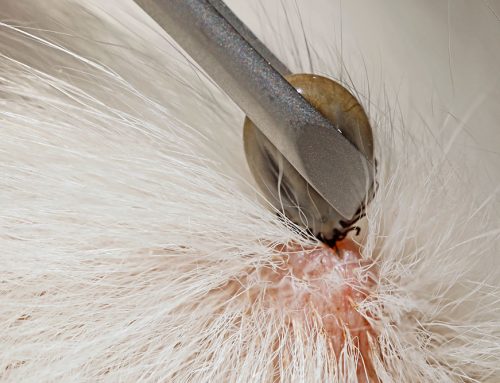
Santa Monica’s active outdoor lifestyle means dogs here encounter a wide variety of environmental exposures, from beach environments to urban parks. Vaccine-site reactions and inflammatory masses from skin trauma or foxtails are more common in active outdoor pets, and understanding whether a mass is reactive and resolving versus growing and persistent guides the testing decision.
Lumps That Should Be Tested Right Away
Some presentations should not be put on a monitoring schedule at all:
- Any mass that has grown noticeably in two to four weeks
- A mass with visible ulceration or bleeding
- Swelling in the jaw, throat, or inguinal region
- Any mass accompanied by enlarged lymph nodes in the same region
- Oral masses of any kind (always warrant prompt evaluation)
- Any new mass in a cat (cats have a higher proportion of malignant masses than dogs)
For these presentations, same-day evaluation is appropriate. If a mass appeared recently and is changing rapidly, urgent care is available during open hours.
What Happens After Results Come Back
Results generally fall into three categories, and each has a clear next step:
Benign results: A monitoring plan. Some benign masses are best left in place with periodic rechecks. Others benefit from eventual removal when they reach a size that causes mechanical problems or start to change.
Malignant results: A direct, honest conversation about options. Surgical excision with appropriate margins, referral to a veterinary oncologist, chemotherapy, radiation, or palliative care, depending on the specific diagnosis, stage, and your family’s goals.
Inconclusive results: Guidance on the next step, whether repeat sampling, biopsy, or imaging to better characterize the mass.
Our team explains findings in plain language and walks through what they mean for your pet’s specific situation.
The Experience for Your Pet
FNA: Brief appointment. The area is clipped or cleaned. The needle is placed over 30 to 60 seconds. Your pet goes home immediately. Results in a few business days.
Biopsy: Pre-anesthetic bloodwork. Procedure 15 to 60 minutes under anesthesia. Same-day discharge in most cases. E-collar at home during healing. Suture removal at 10 to 14 days. Histopathology results in one to two weeks.
Frequently Asked Questions
Can we just monitor a lump and see if it grows?
For some small, likely benign masses, monitoring with specific size-check intervals is appropriate. For rapidly growing, recently changed, oral, or clinically suspicious masses, waiting is not the right call. The discussion at your appointment will clarify which category applies to your pet’s specific lump.
My pet’s lump aspirated as benign last year. Does it need testing again?
Not necessarily, but it should still be measured and monitored. If the mass has grown, changed in texture or appearance, or if new masses have appeared nearby, a repeat aspirate or biopsy is worth doing. For older pets, periodic re-aspiration of long-standing masses is reasonable even without obvious changes.
Will my pet be in pain after biopsy?
Pain management is built into every biopsy procedure, including pre-surgical, intra-surgical, and post-operative oral medication. CO2 laser surgery specifically reduces post-operative discomfort compared to conventional excision, and most pets recover comfortably with appropriate medication.
What if the result comes back malignant?
A malignant result opens a direct, honest conversation about options: surgical excision with appropriate margins, referral to a veterinary oncologist, chemotherapy, radiation, or palliative care, depending on the specific diagnosis, stage, and your family’s goals.
How much does testing cost?
FNA is significantly less expensive than biopsy. Contact us directly to discuss expected costs for your pet’s specific situation before the appointment.
Finding Answers Sooner for Your Pet
The goal of diagnostic testing is not just getting an answer; it is getting the right answer efficiently, so the right treatment can actually work. FNA and biopsy are complementary tools: one fast and preliminary, one definitive. Used together when needed, along with newer screening options, they move you and your pet from uncertainty to a clear plan.
Request an appointment or contact us to have any new or changing mass evaluated by our team.

Leave A Comment